Let’s say you recently went to your primary care physician because of thyroid problems. Your doctor then requested you to undergo a thyroid ultrasound.
Thyroid ultrasound is a technique which employs the use of high frequency sound waves to create images of the thyroid gland. Also called ultrasound scanning or sonography, uses an ultrasound machine, which transmits sound waves to your thyroid gland using an ultrasound transducer. These sound waves travel to your thyroid gland and they hit a boundary between blood and soft tissue or between soft tissue and bone. There are sound waves which get reflected back to the probe and they are then sent back again and back and forth. The ultrasound machine then calculates the distance from the probe to the thyroid gland and its tissues using the speed of sound in tissue (5,005 ft/s or1,540 m/s) and the time of the each echo's return (usually on the order of millionths of a second). The ultrasound equipment then displays the distances and intensities of the echoes on the screen, forming a two-dimensional image. The sound waves are instantly measured and displayed by a computer, which in turn creates a real-time picture on the monitor. One or more frames of the moving pictures are typically captured as still images.
What is interesting is that ultrasound imaging is based on the same principles involved in the sonar used by bats, ships and fishermen. When a sound wave strikes an object, it bounces back, or echoes. By measuring these echo waves it is possible to determine how far away the object is and its size, shape, and consistency (whether the object is solid, filled with fluid or both). In medicine, ultrasound is used to detect changes in appearance of organs, tissues, and vessels or detect abnormal masses, such as tumors.
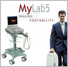
An ultrasound machine looks like this: it usually contains a computer, a video display screen and a probe or transducer which is being placed on the body. The probe is a hand-held device that looks like a microphone attached to a cord. The probe is the one responsible for sending sound waves into the body and picks up the returning echoes from the tissues being examined. The images are then formed on the display monitor and are dependent on the amplitude (strength), frequency and time it takes for the sound signal to return from the patient to the transducer and the type of body structure the sound travels through.
So, why do you think your doctor requested a thyroid ultrasound exam?
Doctors typically request tests which are useful to your diagnosis. A thyroid gland is often imaged under ultrasound to help diagnose a lump in the thyroid or a thyroid which is not functioning properly. Also, it is also used as a guide in procedures such as ultrasound guided needle biopsies. A needle biopsy is a procedure which uses needles to extract sample cells from an abnormal area for laboratory testing. Also, the ultrasound can help guide placement of a catheter or draining device in the thyroid gland.
If indeed, you have been requested to obtain a thyroid ultrasound you should prepare well for the examination. No other special preparation is needed. On the day of the examination, you should wear comfortable, loose fitting clothing for your ultrasound examination. You should also remove all clothing and jewelry in the area to be examined. This is to ensure that there would be no interferences to the test. The technician may also ask you to wear a gown for the procedure.
A pillow will be placed behind the neck to extend the area to be scanned for a thyroid study.
A clear water-soluble
ultrasound gel is applied to the area of the body being studied to help the transducer make secure contact with the body and eliminate air pockets between the transducer and the skin. The sonographer (ultrasound technologist) or physician then presses the transducer firmly against the skin in various locations, sweeping over the area of interest or angling the sound beam from a farther location to better see an area of concern.
There is usually no discomfort from pressure as the transducer is pressed against the area being examined.
If scanning is performed over an area of tenderness, the patient may feel pressure or minor pain from the transducer. The patient will need to extend your neck to gain appropriate access, which may be mildly uncomfortable.
When the examination is complete, the patient may be asked to dress and wait while the ultrasound images are reviewed. However, the sonographer or physician is often able to review the ultrasound images in real-time as they are acquired and the patient can be released immediately. This ultrasound examination is usually completed within 10-15 minutes.
Once the imaging is complete, the gel will be wiped off the skin. The patient is then able to resume normal activities immediately.
Regarding the results, a physician specifically trained to interpret ultrasound examinations, will analyze the images and send a signed report to the patient’s primary care physician or review the results with the patient.
Most ultrasound scanning is noninvasive (no needles or injections) and is usually painless. It is a procedure which is widely available, easy-to-use and less expensive than other imaging methods. It does not use any ionizing radiation. It gives a clear picture of soft tissues that do not show up well on x-ray images. It provides real-time imaging, making it a good tool for guiding minimally invasive procedures such as needle biopsies and needle aspiration.
